
03 CERVICAL VERTEBRAE
Typical cervical vertebrae
There are seven cervical vertebrae. C3–C5 are "typical" and largely interchangeable:
- Small kidney-shaped body,
- Large triangular vertebral foramen (accommodating the cervical enlargement of the cord),
- Short bifid anteriorly-tilted spinous process, that accommodate the ligamentum nuchae,
- Transverse foramen transmitting the vertebral artery, vein and sympathetic plexus, and
- Uncinate processes on the superolateral body forming the uncovertebral joints of Luschka, a common site of osteophyte formation causing cervical radiculopathy.
Atypical cervical vertebrae
C1, Atlas
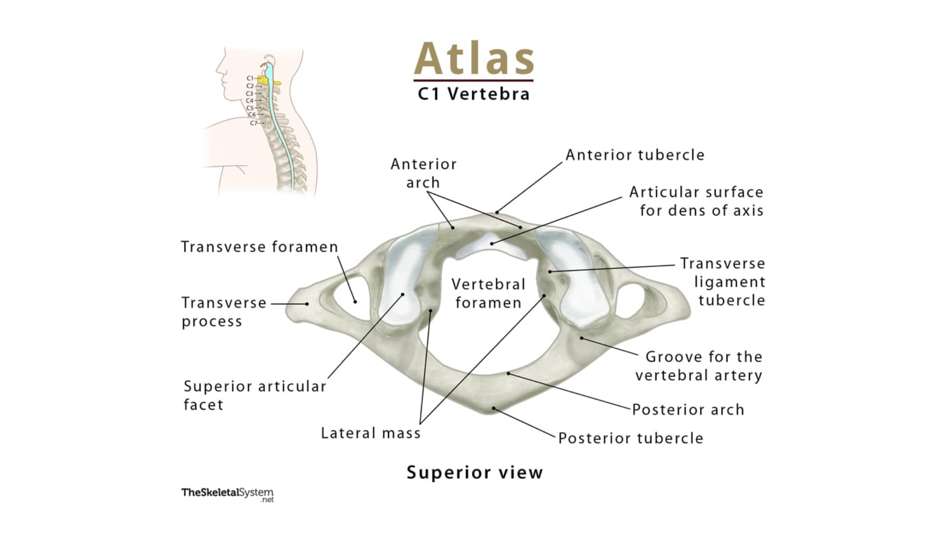
No body and no spinous process, just an anterior and posterior arch united by lateral masses; essentially a bony ring that cradles the occipital condyles, allowing the "yes" nod at the atlanto-occipital joint.
👩⚕️ Jefferson's fracture is a burst fracture of the C1 ring from axial loading (diving into shallow water, helmet impact).
C2, Axis
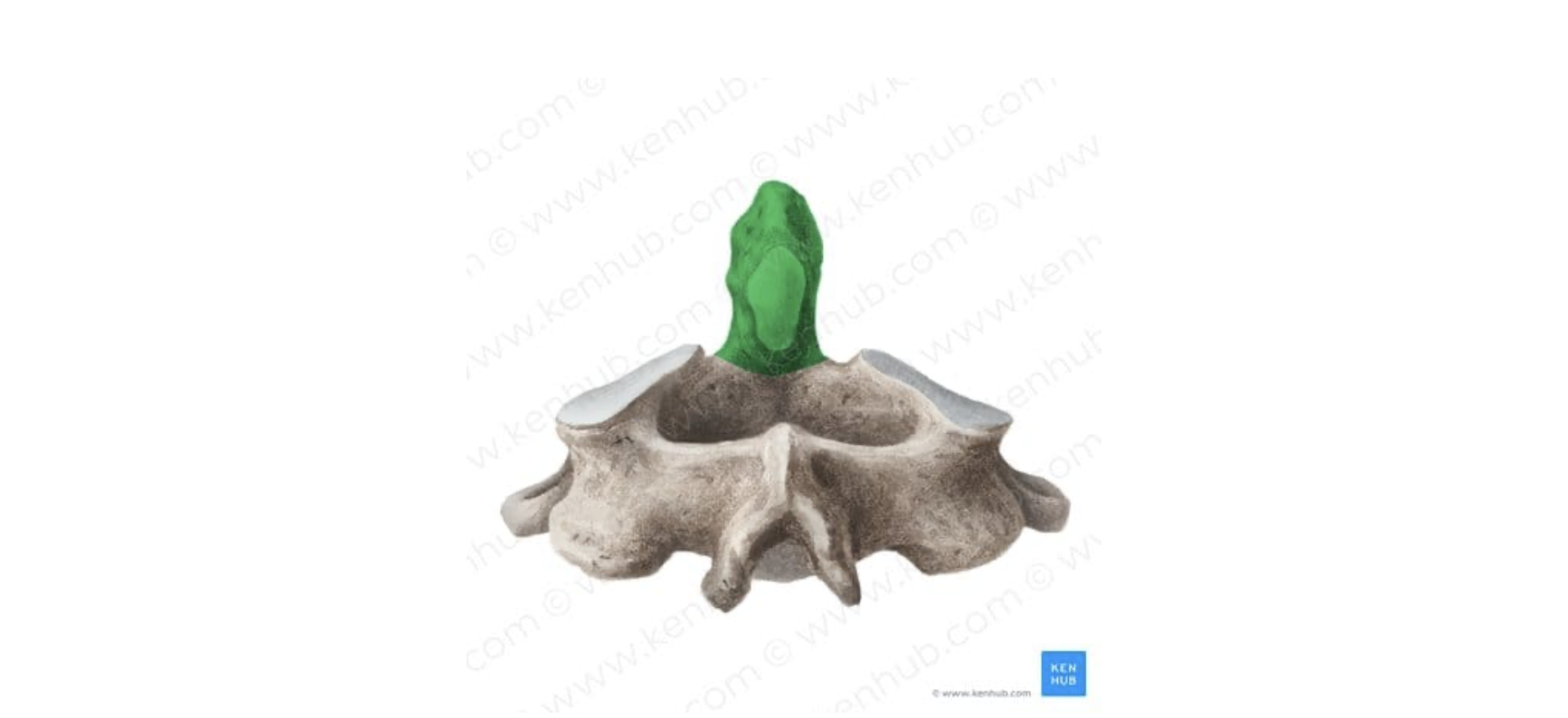
Long bifid spinous process plus the odontoid peg (dens) projecting upward from its body. The dens is held against the anterior arch of C1 by the transverse ligament of the atlas, forming a pivot that produces the "no" shake at the atlanto-axial joint.
👩⚕️ Hangman's fracture is a bilateral pars interarticularis fracture of C2, classically from hyperextension
C6
C6 has a prominent carotid (Chassaignac's) tubercle on the anterior tubercle of the transverse process. Four high-yield facts cluster here:
- The vertebral artery enters the transverse foramen at C6 (it skips C7's foramen) and ascends through C5–C1 to enter the skull via the foramen magnum.
- The common carotid artery can be compressed against the carotid tubercle — useful for haemorrhage control and historically for diagnosing carotid pathology.
- "C6 is where everything begins": The trachea (larynx becomes trachea), oesophagus (pharynx becomes the oesophagus), recurrent laryngeal nerve all start at C6.
- The cricoid cartilage lies at the level of C6. Cricothyroidotomy is performed through the cricothyroid membrane just above the cricoid.
C7, Vertebra prominens
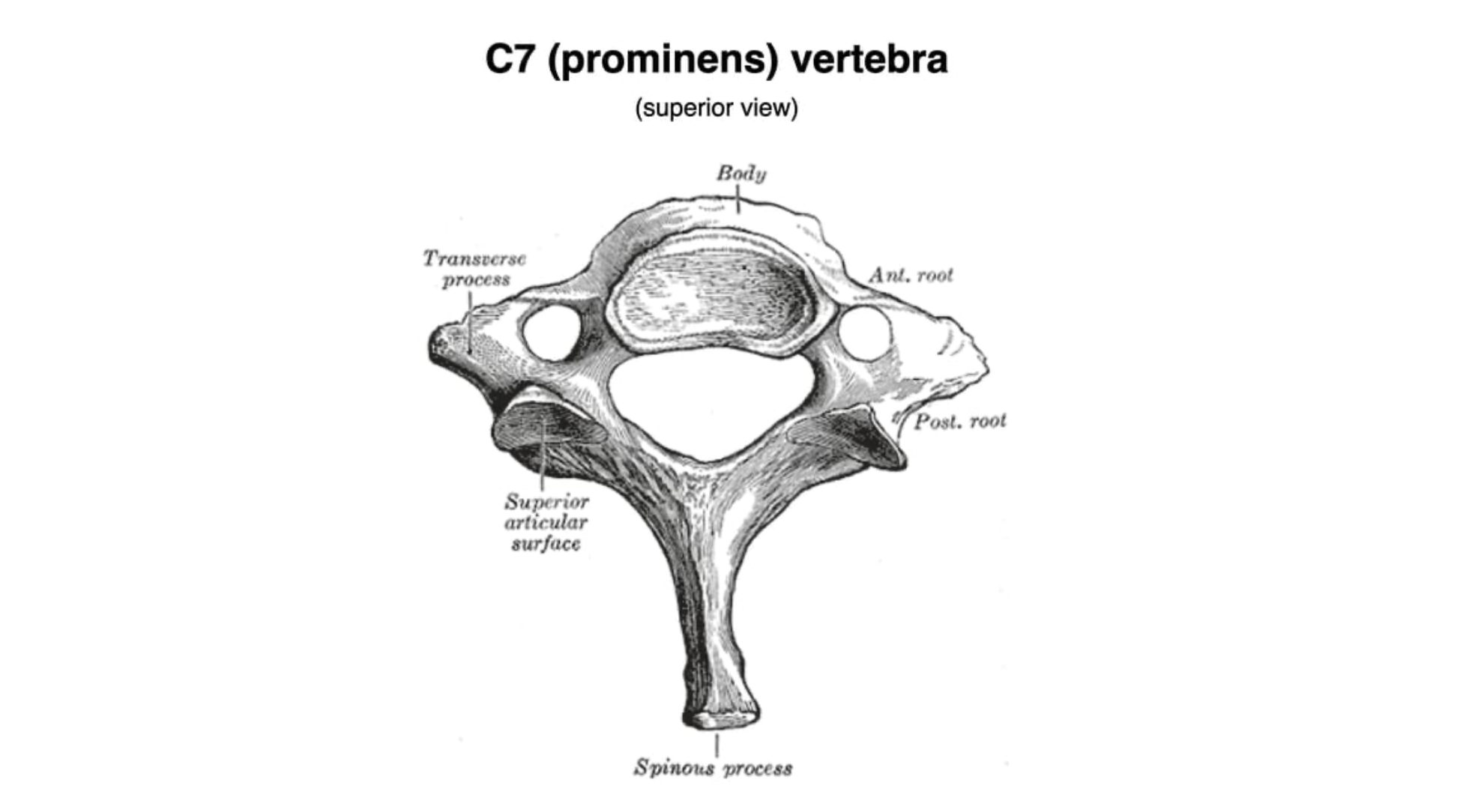
Long, non-bifid spinous process — the most palpable vertebral landmark at the base of the neck. Its transverse foramen, when present, transmits only small accessory veins, not the vertebral artery.

Which cervical vertebra is characterised by a long bifid spinous process?
- ((C1 (Atlas)::No spinous process — just anterior and posterior arches around the dens.))
- ((C2 (Axis)::☑️ Long bifid spinous process plus the odontoid peg (dens).))
- ((C6::Has the anterior carotid tubercle, not a bifid spine.))
- ((C7 (Vertebra prominens)::Long spinous process but non-bifid — most palpable posteriorly.))
The vertebral artery enters the foramen transversarium at which level?
- ((C4::Level of carotid bifurcation, not vertebral artery entry.))
- ((C5::Vertebral artery has already entered one level lower.))
- ((C6::☑️ Enters at C6, marked by the prominent carotid (Chassaignac's) tubercle.))
- ((C7::Transverse foramen of C7 transmits only small accessory veins, not the artery.))
👩⚕️ The vertebral artery skips C7 — a classic SBA trap.
At which vertebral level can the common carotid artery pulsation be compressed, and the trachea begin?
- ((C4::Level of carotid bifurcation, not tracheal start.))
- ((C5::Mid-thyroid cartilage — no major landmark begins here.))
- ((C6::☑️ Common carotid compressed against Chassaignac's tubercle; cricoid, trachea and oesophagus all begin.))
- ((T4/T5::Sternal angle — site of tracheal bifurcation, not its start.))
Which cervical vertebra is most prominently felt on the back of the neck?
- ((C5::Not typically palpable.))
- ((C6::Level of cricoid, palpable anteriorly but not the posterior prominence.))
- ((C7::☑️ Vertebra prominens — long, non-bifid spinous process, easily palpable.))
- ((T1::Sometimes palpable but C7 dominates the posterior landmark.))